
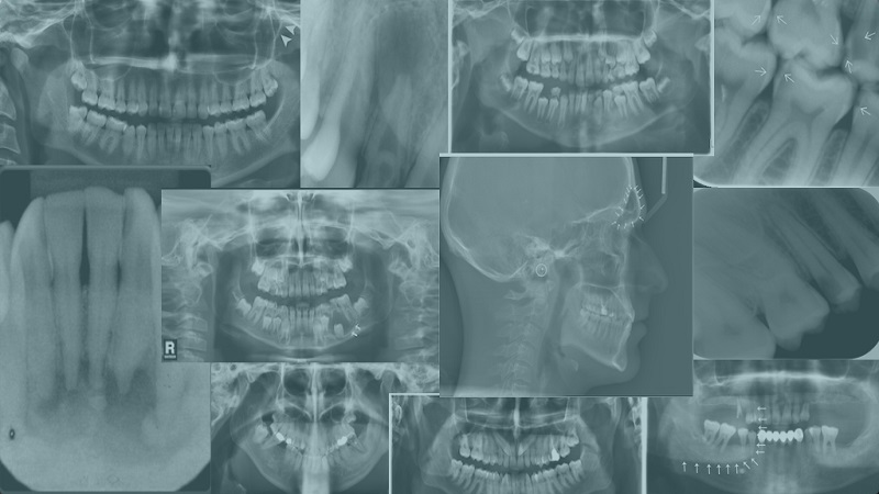
Getting to know the types of X-ray images of the mouth
In this article, the website of Dr. Hossein Borjian The best gum surgeon in Isfahan We will examine the types of X-ray images of the mouth. There are different types of oral radiology photos that have differences from each other. The most common types of photos X-Ray Inside the mouth are as follows:
Intraoral X-rays
Byte Wing :
In this technique, you bite a piece of paper so that the dentist can see the state of contact between the crowns of the teeth. This photo is mostly used to check caries between the teeth.
occlusal:
In this x-ray, you close the jaws so that the upper and lower teeth are aligned and the dentist checks their contact with each other.. In this radiology photo, you can also identify anatomical abnormalities of the floor of the mouth or the roof of the mouth.
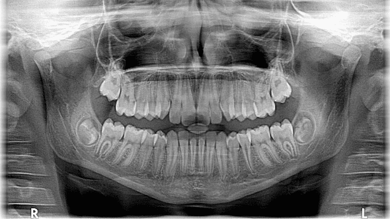
panoramic:
In this method of photographing the mouth, the device rotates around your head and takes pictures. The dentist uses this photo to check the condition of the back teeth, planning for implantation Dental implant or jaw problems uses.
Periapical:
A periapical photograph focuses on two complete teeth from root to crown.
Suggested content : Disadvantages and advantages of invisible orthodontics
Extraoral x-rays
X-ray image from outside the mouth if the Isfahan dentist suspects problems in the areas outside the gums and teeth. (For example, the jaw) It is true, it is done.
Learn more about maxillofacial surgery.
Panoramic photo:
It shows the entire range of the mouth. And all upper and lower teeth can be seen in only one photo. This photo shows the position of erupting and erupting teeth, and it shows impacted teeth, tumors, and more..
Tomography:
In the tomography, a certain layer or part of the mouth can be seen and the other layers are blurred. This photo shows structures that are difficult to see clearly because they are adjacent to other structures and are obstructed by other structures..
Cephalometric:
In the cephalometric photo, one side of the head can be seen. In this photo, the position of the teeth in relation to the jaw and facial profile is determined. The orthodontist uses the cephalometric photo for treatment planning..
Sialography:
In the sialography photo, a special dye is used that is injected into the salivary glands so that it can be seen better in the X-ray photo.. Salivary glands are soft tissues that cannot be seen on regular x-rays. The dentist will order this photo to check for problems with the salivary glands, such as blocked salivary glands or Sjogren's syndrome.
CT Scan:
CT scan or computed tomography is a type of imaging. in which the internal structures are seen in three dimensions. This imaging is used to identify facial bone problems such as cysts, fractures, and tumors.
Cone beam CT:
In this type of X-ray, three-dimensional images of facial structures, soft tissues, nerves and bones are obtained.. These photos help the dentist to determine the right place for implant placement and show oral and facial cysts or tumors.. Also, this photography is used to check the problems of gums, roots of teeth and jaws. Cone beam CT is similar to a regular CT scan. Both methods provide high-quality images, but the way they take pictures is different.
The Instagram page of Dr. Hossein Borjian, the best gum surgeon in Isfahan
The cone beam CT machine rotates around the patient's head. And it gets all the information in just one turn. In a normal CT scan, the device rotates several times around the patient's head and collects information layer by layer. This imaging method exposes the patient to a lot of radiation. The unique advantage of cone beam CT is that it can be used in the dentist's office. CT scan equipment is available only in imaging centers or hospitals.
Digital photography:
This x-ray sends two-dimensional images of the mouth directly to the computer. Photos are viewed and saved on the computer screen and printed with a printer within a few seconds. Digital imaging has several advantages over other x-ray imaging methods. These photos can be enlarged and seen more clearly. It is also easier for the dentist to observe the smallest changes that cannot be detected in the examination in these images. If necessary, the dentist can immediately send the photo to another colleague through the Internet to ask for his opinion. The amount of radiation in a digital photo is less.
Ways of communication with the specialized dental center of Najm
Attention :
- The scientific accuracy of the above published material should be confirmed by the patient's personal consultation with Dr. Borjian.
- This article is managed and published by the site admin.