

Familiarity with CBCT images for dental implants
Dental CBCT image is very important for implants, bridges and deep restoration of damaged oral and dental tissues. of the year 1960 Since the implant was introduced as one of the methods of restoring damaged teeth, the photos taken of the jaw and teeth with traditional x-rays have always been an inevitable and necessary thing until today's advanced technology.. In this article from Dr. Hossein Borjian's website The best gum surgeon in Isfahan We examine the familiarity with CBCT image for dental implants.
Today, the world of various oral and dental prostheses such as implants, bridges, etc… They are still evolving and improving. By using CBCT imaging, the installation process and efficiency of these parts can be done more accurately and efficiently. For this reason, dental teams use dental CBCT images for implants and other methods of repairing damaged tissues to accurately check the condition of the teeth, the thickness of the jaw bone, and other factors..
What is a dental CBCT image?
Dental CBCT, or cone beam computed tomography, is an advanced imaging technology that provides a three-dimensional view (3D) It presents a joint of the jaws, teeth and surrounding structures to the specialist doctor. This information helps dentists and oral and maxillofacial surgeons to place dental implants more accurately and reliably inside the gum and oral space..
There are several reasons why dentists use dental CBCT scans for their diagnosis and treatment recommendations. In other words, the best course of action when planning a dental implant for a patient is to use a dental CBCT scan. As we know, dental implants consist of two components, one of which is a titanium base that is placed in the jawbone and acts as an anchor for the dental implant.. However, in order for the implant to be placed safely and without disturbing vital nerves or other important structures, it is essential that the dentist can see its placement and accurately examine the intended space; This is where a CBCT scan comes in.
CBCT scan structure
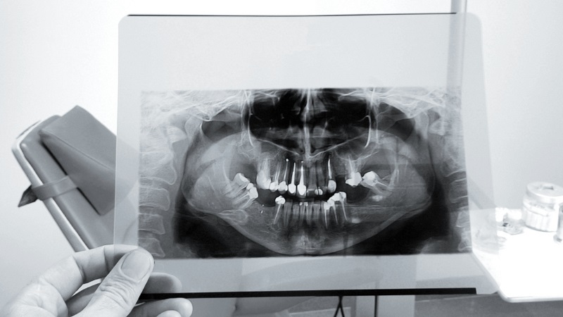
A CBCT scan produces three-dimensional cross-sectional images of the jaws and teeth that provide detailed information about the area of interest.. This information cannot be obtained from conventional radiology and conventional x-rays. Dental CBCT imaging allows the dentist to evaluate and check the location of the implant post. During this examination, he can determine that the bone has the right shape and density to grow back around the implant and secure it in place.. Also, the use of this technology minimizes the risk of problems such as nerve damage and makes the whole process safer and increases the probability of successful application of different prostheses..
Applications of CBCT scanning for dentists
The advantages of cone beam CT scanning make it an excellent choice for dentistry and implant placement. Using this method can have many benefits for safe treatment programs such as implants.
- Using a dental CBCT image for an implant makes it easy for the dentist to measure and localize the existing jawbone. This allows virtual implant placement to be performed with complete precision.
- Dentists can use CBCT and an optical scan to create a complete virtual model of a patient's soft tissues, bones, and teeth..
- With the help of CBCT scan, the dentist can design the right bite for the patient's teeth and reduce the risk of inconsistency of implants..
- The dentist uses the CBCT scan to determine the location of the sensory nerves to choose the appropriate length of the implant and reduce the risk of nerve damage.
- CBCT imaging provides the dentist with an accurate picture of the location of the maxillary sinus. In this way, the surgeon can choose the appropriate length of the implant to prevent the penetration of the implant into the sinus.
- Cone CT scans allow the dentist to select the appropriate implant size for optimal stability and integrity.. This ensures that the implant can meet the patient's needs and serve the individual for a long time.
Comparison of CBCT scan and full mouth or panoramic imaging
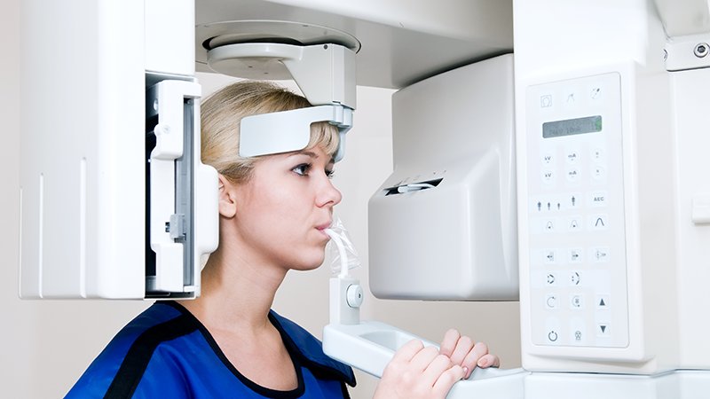
CBCT stands for Cone Beam Computed Tomography. which are used by dental professionals. This system rotates around you and uses cone-shaped x-rays to record the state of your mouth and teeth or other data.. With the help of CBCT imaging in less than one minute, approx 150-200 The image is taken from different angles. Data taken to reconstruct a 3D image of different parts of your body such as teeth, mouth, jaw and neck, ears, throat and nose (ENT)Taken. Probably already about full mouth x-rays (FMX) or panoramic x-rays (HERE) You have heard from your dentist. FMX usually every 3-5 Sal is recommended by your dentist and takes pictures of each tooth and its surrounding structures. This technology is used to diagnose gum disease, cavities, dental abscesses and lesions.
The Instagram page of Dr. Hossein Borjian, the best gum surgeon in Isfahan
PANO technology can also produce a single image, which is often used by oral surgeons and orthodontists. Used for superficial diagnosis of oral and dental diseases. This type of X-ray does not provide clear details, so it cannot be relied on for deeper and more accurate diagnosis and investigations.. Compared to these standard x-rays, a dental CBCT scan is a much more accurate and effective way to get information about your dental health.. CBCT technology also uses less radiation, requiring fewer scans to see different views and angles of the mouth.. Additionally, unlike traditional dental X-rays, a cone beam CT scan can show both bone and soft tissue..
Attention :
- The scientific accuracy of the above material should be consulted with the patient in person with Mr. Dr. Berjian, Mastership Gum and bone grafting be confirmed.
- This article was managed and published by the site admin.
Read more :
Special anesthetic drugs in dentistry
Symptoms of wisdom tooth extraction