
Examining the uses of OPG photos
In the rest of this article from Dr. Hossein Borjian's website The best dentist in Isfahan We introduce you to the uses of OPG photos. X-ray imaging has many uses in the medical world. X-rays in dentistry (Radiology) It is taken in different ways. Panoramic and periapical radiology images are two-dimensional images taken from three-dimensional objects. Also the photo of OPG (OPG) Teeth and cephalography also have many uses.
OPG photo definition
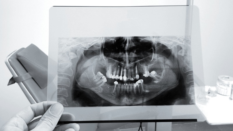
His photo is PG (OPG) Abbreviation of orthopantomonogram (Orthopantomogram) It is a kind of X-ray photo of the teeth. The photo of PG has a panoramic view of the jaw. While the cephalogram photo is a photo X-Ray It is one of the structures of the face and the normal dental radiology photo is also a photo. which is taken from the structure of one or more teeth and a little from the tissues around it (which itself has different types).
Suggested content : Dental orthodontic procedures
Applications of OPG photo
The uses of OPG photos are as follows:
- Examining the general position of the teeth and their relationship with each other
- decay
- Tooth impaction
- Fracture or cracking
- Discoloration of teeth
- infection
- tumor
- The condition of the sinuses
- The condition of the jaw bone
- Dental implant placement
- Orthodontic treatment
- Chin and jaw surgery
Complications of OPG photo
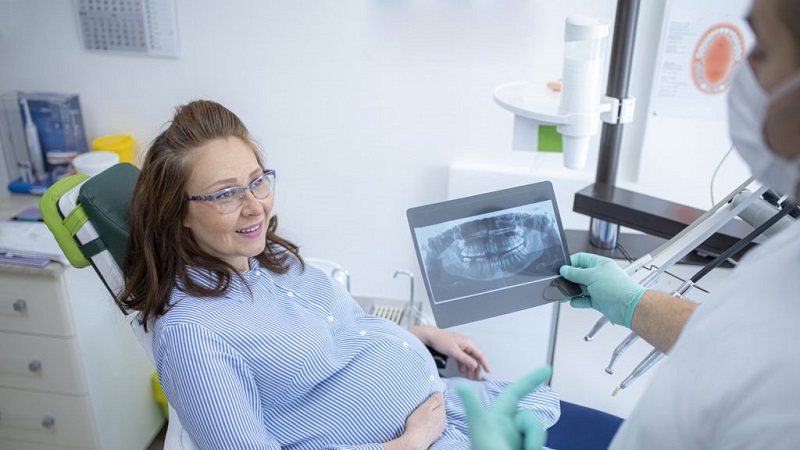
To take this type of dental photo, you need to be exposed to a small amount of radiation. The radiation dose is not high and there is nothing to worry about. If you are pregnant, you should inform the dentist.
Benefits of OPG photo
- This photography method is immediate and completely painless. By taking an opg photo, very good information is provided to the dentist, orthodontist or maxillofacial surgeon.
- In this photo, all upper and lower teeth can be seen in a single image.
- In this image, the number of teeth, their position and angle, and any teeth hidden under the gums or extra can be seen.. For this reason, it is necessary for orthodontic treatment.
- These images are needed to examine the condition of the wisdom teeth and the structure around them and to decide whether to extract them or not.
- To implant a dental implant, the dentist needs to observe the overall structure of the mouth and teeth and their position.
The Instagram page of Dr. Hossein Borjian, the best dentist in Isfahan
Comparison of OPG image with conventional dental radiology image
In an OPG image, all the teeth and the jawbone around them are seen in one image, but in a conventional radiology image, only one or more teeth are seen from a close angle.. Although this is to see a more precise condition of a specific tooth (For example, the condition of the tooth pulp or damage to the nerves inside it or possible small cracks) It is more useful, but it is not suitable for seeing the general view of the teeth. With the OPG photo, any problem can be seen anywhere in the mouth. Also, problems related to the jaw bone and its relationship with the head and temporomandibular joint (jaw joint) Characterized. Sometimes both types of X-rays may need to be taken.
Ways of communication with the specialized dental center of Najm
Attention :
- The scientific accuracy of the above published material should be confirmed by the patient's personal consultation with Dr. Borjian.
- This article is managed and published by the site admin.